
Furthermore, the actin waves promote the formation of more microtubule filaments. When a wave of actin reaches a projection, the projection grows for a while and then stops until the next actin wave arrives. The experiments show that a protein called actin forms a mesh of filaments in a wave-like manner, starting in the cell body and moving outwards into the projections. used microscopy to study the transport of axon-promoting proteins in hippocampal neurons.

However, it is not clear what drives these fluctuations. In immature neurons, kinesin motors randomly move in and out of different projections, before settling in the projection that will grow into the axon. These axon-promoting proteins are carried to the axons by a motor protein called kinesin, which moves along fibers called microtubules. It is believed that signal proteins inside the neuron that promote the formation of an axon selectively accumulate in a projection as it grows into an axon. These projections randomly retract and lengthen for a while before a single projection grows into an axon and the others become dendrites. In the early stages of development, an immature neuron sends out multiple projections that extend out in all directions from its cell body.
#NEURITE DENDRITE AXON SERIES#
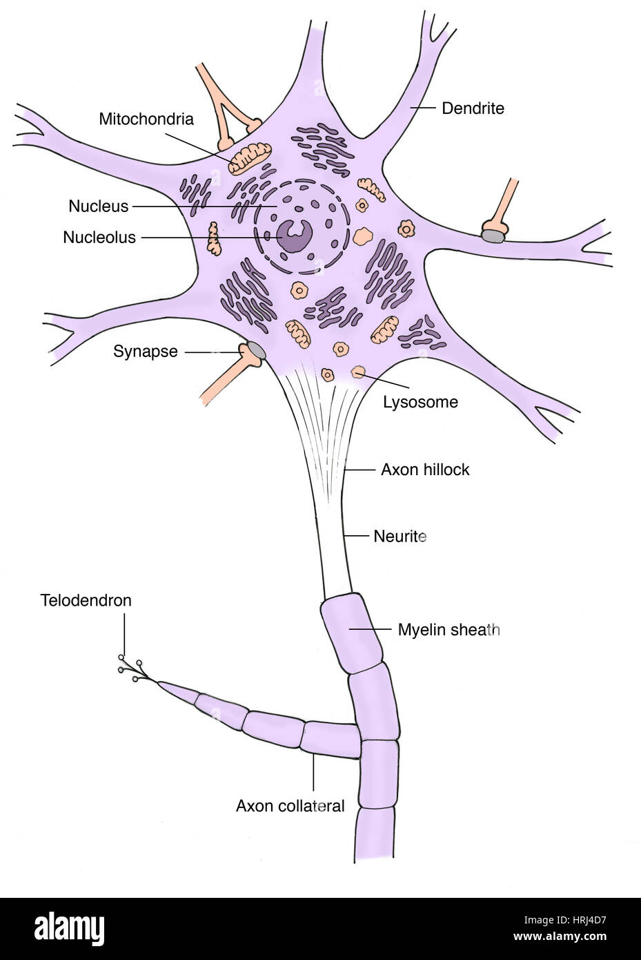
In animal embryos, immature neurons in part of the brain called the hippocampus – which is crucial for learning and forming memories – develop into mature neurons through a series of steps. Signals are received by branch-like projections called dendrites, pass through the cell body and then pass along a long projection called the axon before being transmitted to the dendrites of neighboring neurons. Nerve cells (also known as neurons) connect with each other to form complex networks through which signals are carried around the body. We propose that actin waves create large stochastic fluctuations in microtubule-based transport and neurite outgrowth, promoting competition between neurites as they explore the environment until sufficient external cues can direct one to become the axon. Actin waves also require microtubule polymerization, arguing that positive feedback links these two components. Our data argue for a mechanical control system whereby actin waves transiently widen the neurite shaft to allow increased microtubule polymerization to direct Kinesin-based transport and create bursts of neurite extension. We show that actin waves, which stochastically migrate from the cell body towards neurite tips, direct microtubule-based transport during the multi-polar state. Hallmarks of the multi-polar state are large fluctuations in microtubule-based transport into and outgrowth of different neurites, although what drives these fluctuations remains elusive. Generally, target detection can be accomplished using chromogenic or fluorescent staining.Many developing neurons transition through a multi-polar state with many competing neurites before assuming a unipolar state with one axon and multiple dendrites. Immunolabeling of cells (immunocytochemistry or ICC) or tissues ( immunohistochemistry or IHC) with antibodies to study neurons is a highly utilized application in Neuroscience mainly due to the availability of a wide range of markers and the relatively low cost for performing and imaging the immunolabeled material. Measurement of protein expression levelsĭownload our complimentary Neuronal Cell Markers Poster.Assessment of cellular or protein co-localization.Phenotypic and morphological analysis of neurons.microglia, astrocytes, and oligodendrocytes) Distinguishing neurons from other cell types in the nervous system (e.g.The use of antibodies for these markers in conjunction with microscopy serves as a powerful method for: These unique compartments are distinguishable using specific markers, and are generally classified into soma (cell body), axon, dendrite, and synapse. Neurons have highly compartmentalized structures that allow their distinction from other cell types in the nervous system.


 0 kommentar(er)
